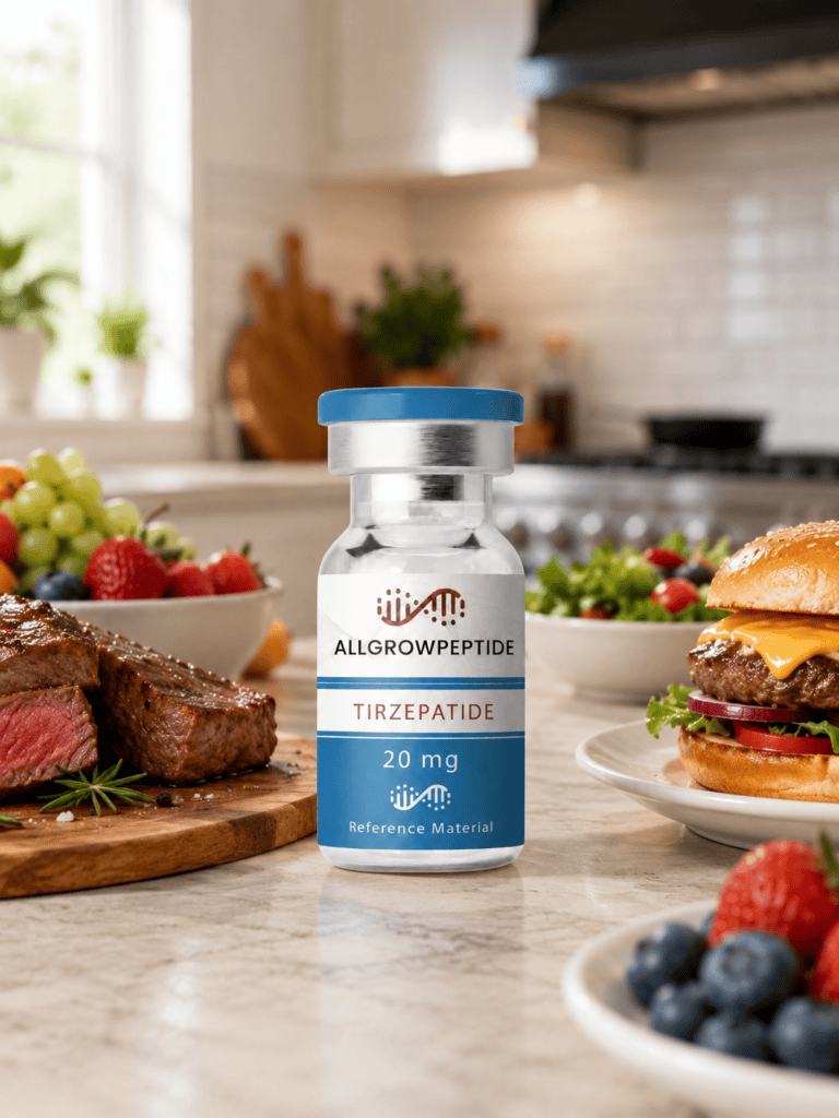
Retatrutide 10mg (Rt10)has become an increasingly visible term in peptide research catalogs. However, the “10mg” designation refers to a material quantity or vial presentation. It does not define a different molecular species.
The molecule itself is Retatrutide, also known by the research identifier LY3437943. It belongs to a class of engineered peptide agonists designed to interact with several metabolic G protein-coupled receptors.
Researchers study Retatrutide because one molecule can activate three receptor systems. These are the glucose-dependent insulinotropic polypeptide receptor, the GLP-1 receptor, and the glucagon receptor. Published work therefore describes LY3437943 as a triple agonist peptide.
This article examines Retatrutide from a molecular biology and laboratory perspective. It covers structure, receptor signaling, biochemical mechanisms, comparison with related peptides, analytical verification, stability, and practical research considerations.
For Research Use Only. Not for human use.
What Is Retatrutide 10mg?
Retatrutide is the international name commonly used for LY3437943.
It is a synthetic, chemically modified peptide rather than a protein, vitamin, nucleotide, or conventional small molecule.
Published research describes Retatrutide as a 39-amino-acid peptide engineered to activate three class B G protein-coupled receptors:
GIP receptor, or GIPR
GLP-1 receptor, or GLP-1R
glucagon receptor, or GCGR
The reported molecular formula is C221H342N46O68. The molecular mass is approximately 4731.33 Da.
The peptide also contains chemical modifications that distinguish it from naturally occurring metabolic hormones.
One important modification includes a C20 fatty diacid moiety. This structural feature promotes albumin association and alters pharmacokinetic behavior in experimental systems.
That classification is more accurate than informal terms such as “GLP-3.”
Lilly specifically states that “GLP-3” is not a valid scientific classification. Instead, “triple agonist” describes the mechanism more accurately.
Practical Experimental Considerations: What Retatrutide Is — and Is Not
Retatrutide is a peptide.
However, researchers should not treat it as an endogenous peptide hormone.
Its sequence contains deliberate structural modifications. Therefore, its physicochemical and receptor properties differ from native GIP, GLP-1, and glucagon.
It is also not a mixture of three hormones.
Instead, one engineered peptide molecule interacts with three receptor classes.
This distinction matters during experimental design.
A laboratory comparing Retatrutide with native GLP-1 should not assume equivalent potency, receptor selectivity, degradation kinetics, or assay behavior.
Likewise, “Retatrutide 10mg” does not describe a special molecular variant.
The mass simply describes the nominal amount of research material in a particular presentation.
Retatrutide contains 39 amino-acid positions and several engineered features.
The molecule incorporates non-standard amino-acid chemistry. It also includes a lipid-linked side chain.
Consequently, researchers should view Retatrutide as a chemically modified peptide rather than a simple linear chain of natural amino acids.
The lipid modification has an important biochemical purpose.
A C20 fatty diacid supports reversible albumin association. Consequently, the peptide behaves differently from short, unmodified peptide ligands in biological matrices.
Peptide bonds still form the main backbone.
However, side-chain modifications add additional structural complexity.
This complexity affects:
reversed-phase chromatography;
mass-spectrometric ionization;
solubility;
aggregation behavior;
nonspecific surface binding;
receptor interaction;
apparent stability.
Therefore, laboratories should avoid applying analytical assumptions developed for small, hydrophilic peptides without validation.
Major Research Pathways and Mechanisms
GIP Receptor Signaling
Starting trigger: Retatrutide interacts with GIPR.
GIPR belongs to the class B family of G protein-coupled receptors.
After ligand binding, the receptor commonly couples to Gs proteins.
This activates adenylyl cyclase.
Adenylyl cyclase then increases intracellular cyclic AMP, or cAMP.
Consequently, downstream signaling may involve protein kinase A and other cAMP-sensitive pathways.
Researchers can investigate this pathway through:
cAMP accumulation assays;
receptor binding studies;
reporter-gene systems;
receptor internalization assays;
β-arrestin studies.
Retatrutide shows relatively strong activity at GIPR compared with the corresponding endogenous ligand profile described in its discovery research.
GLP-1 Receptor Signaling
Starting trigger: Retatrutide binds GLP-1R.
Like GIPR, GLP-1R belongs to the secretin-like class B GPCR family.
Ligand binding stabilizes an active receptor conformation.
The receptor then couples primarily to Gs signaling.
This process increases intracellular cAMP.
Researchers can measure downstream events with cell-based functional assays.
Structural biology now provides direct evidence for Retatrutide interaction with GLP-1R.
A deposited cryo-electron microscopy structure captured Retatrutide bound to human GLP-1R in a G-protein complex at approximately 2.68 Å resolution.
This structural information can support:
molecular docking validation;
ligand-receptor interaction studies;
mutagenesis experiments;
receptor activation models;
structure-activity relationship research.
Glucagon Receptor Signaling
The third major target is GCGR.
This receptor also belongs to the class B GPCR group.
Activation can stimulate Gs signaling and cAMP production.
Researchers often study GCGR alongside metabolic signaling, hepatic cell models, and ligand selectivity.
A Retatrutide-bound human GCGR-Gs cryo-EM structure has also been deposited.
That complex reached approximately 2.84 Å resolution.
Therefore, laboratories can now compare experimentally determined receptor complexes rather than relying only on computational models.
Why Triple Agonism Matters in Experimental Biology
RT10 peptide injection
A single receptor assay answers a relatively narrow question.
A triple agonist creates a more complex experimental system.
Retatrutide allows researchers to examine how receptor activities interact.
For example, a project may compare:
GLP-1R-only signaling;
GIPR plus GLP-1R signaling;
GIPR, GLP-1R, and GCGR signaling.
This design can help separate pathway-specific effects from combined receptor activity.
Researchers may also examine receptor bias.
For example, one ligand might generate strong cAMP signaling but weaker β-arrestin recruitment.
Another ligand may show a different profile.
Therefore, functional assays should not rely on receptor-binding data alone.
Retatrutide, tirzepatide, and semaglutide all contain peptide backbones.
However, their receptor profiles differ substantially.
Semaglutide primarily targets GLP-1R.
Tirzepatide targets GIPR and GLP-1R.
Retatrutide adds GCGR activity to the experimental profile.
Therefore, laboratories can use these compounds in comparative receptor studies.
Such experiments may examine:
potency;
receptor selectivity;
cAMP production;
receptor trafficking;
ligand-induced internalization;
cellular signaling;
structure-activity relationships.
Researchers should still validate each compound independently.
Similar catalog placement does not establish analytical equivalence.
Retatrutide vs Tirzepatide: Scientific Comparison
FDA documentation describes tirzepatide as a 39-amino-acid peptide with a molecular mass of 4813.53 Da. It contains a C20 fatty diacid modification.
Consequently, these molecules share important design concepts.
However, they should not be considered interchangeable.
Their amino-acid sequences and receptor pharmacology differ.
How Retatrutide Is Studied in the Laboratory
Researchers can investigate LY3437943 peptide activity using several experimental systems.
Receptor-expressing cell lines
Engineered cells can express GLP-1R, GIPR, or GCGR separately.
Researchers can then measure receptor-specific activity.
This approach helps isolate one signaling pathway.
cAMP assays
Because all three receptors can couple to Gs proteins, cAMP provides a useful functional readout.
However, cAMP alone cannot describe every signaling event.
Additional assays may provide complementary information.
β-Arrestin and receptor trafficking assays
These experiments can evaluate receptor desensitization, recruitment, and internalization.
They may also reveal signaling bias.
Structural biology
Cryo-EM data provide direct structural information for receptor-ligand complexes.
Researchers can combine these structures with mutagenesis or computational modeling.
Stability and Laboratory Handling
Retatrutide stability depends on formulation, concentration, container, buffer, temperature, and storage duration.
Therefore, one universal storage specification should not be presented as experimentally proven for every research preparation.
For lyophilized research material, laboratories commonly favor frozen, dry, light-protected storage.
However, researchers should follow batch-specific stability documentation whenever available.
Lyophilized material
Keep the vial tightly sealed.
Limit moisture exposure.
Avoid repeated warming and cooling.
In addition, record the initial storage date.
Reconstituted research solutions
Solutions generally require more careful stability control than dry material.
Proteolysis is not the only concern.
Chemical degradation may also occur.
Possible mechanisms include:
oxidation;
deamidation;
hydrolysis;
aggregation;
adsorption to container surfaces.
Therefore, laboratories should validate solution stability under their own buffer conditions.
pH considerations
No single formulation-independent optimal pH should be assumed.
Instead, researchers should evaluate peptide integrity across the buffer range required by the assay.
HPLC or LC-MS can support such stability studies.
Maintaining Sample Integrity
Peptide Purity Selection
1. Allow Controlled Temperature Equilibration
Timing: Several minutes may be appropriate, depending on vial size.
Allow a closed vial to equilibrate before opening.
This reduces condensation risk.
A common mistake is opening a very cold vial immediately.
2. Inspect the Container
Check the vial before preparation.
Look for seal damage, unexpected moisture, or visible material changes.
Record the batch number.
3. Prepare the Experimental Solvent
Use a solvent or buffer compatible with the intended assay.
Researchers should validate pH and ionic strength.
Do not assume one solvent suits every experiment.
4. Reconstitute Carefully
Add the selected research solvent using an established laboratory SOP.
Avoid unnecessary foaming.
Strong agitation may increase aggregation risk for some peptides.
5. Confirm Complete Dissolution
Inspect the solution.
However, visual clarity does not prove molecular integrity.
Use analytical testing when experimental accuracy requires it.
6. Produce Aliquots
Create working aliquots when repeated experiments are planned.
This reduces unnecessary freeze-thaw cycles.
7. Record Preparation Details
Document:
concentration;
buffer;
date;
lot;
storage condition.
Good records support reproducibility.
8. Verify Long-Term Integrity
For extended studies, compare stored material with a reference time point.
HPLC and LC-MS can reveal emerging degradation products.
Analytical and Quality Considerations
HPLC
Reverse-phase HPLC can separate Retatrutide from many related impurities.
Therefore, it is useful for chromatographic purity assessment.
However, an HPLC area percentage alone does not prove molecular identity.
LC-MS
Liquid chromatography-mass spectrometry combines chromatographic separation with mass detection.
It can support molecular identity and reveal mass-shifted impurities.
Tandem MS
MS/MS can provide additional structural evidence.
Fragmentation patterns help confirm peptide sequence regions.
FDA quality documentation for the structurally related peptide tirzepatide describes LC-MS, LC-MS/MS, peptide mapping, and circular dichroism as characterization tools.
Concentration testing
Purity and concentration are different properties.
A sample may show high chromatographic purity yet contain a different total amount than expected.
Therefore, laboratories should use an appropriate quantitative method when absolute concentration matters.
Frequently Asked Questions About Retatrutide 10mg
What is Retatrutide 10mg?
Retatrutide 10mg generally describes a research presentation containing a nominal 10 mg quantity of Retatrutide. The molecule is also known as LY3437943. Retatrutide is a synthetic modified peptide designed to activate GIPR, GLP-1R, and GCGR. The 10mg designation does not create a separate chemical form. Researchers should therefore record the compound identity, lot number, actual assay value, and analytical documentation separately.
Is Retatrutide a peptide?
Yes. Retatrutide is a chemically modified 39-amino-acid peptide. It contains peptide bonds and engineered structural modifications. Its design includes a C20 fatty diacid component associated with altered pharmacokinetic behavior.
What is the molecular weight of Retatrutide?
The reported molecular weight is approximately 4731.33 Da. Database records also report the molecular formula C221H342N46O68. Researchers should distinguish the theoretical molecular mass from measured mass-spectrometric data generated for a specific sample.
Why do researchers study Retatrutide?
Researchers study Retatrutide because it combines agonist activity at GIPR, GLP-1R, and GCGR within one molecule. This feature enables multi-receptor signaling studies. It also supports comparative work involving single-, dual-, and triple-receptor peptide systems.
What is the difference between Retatrutide and Tirzepatide?
The major mechanistic difference concerns receptor targeting. Tirzepatide activates GIPR and GLP-1R. Retatrutide also activates GCGR. Both are modified 39-amino-acid peptides, but their sequences and pharmacological profiles differ.
What does RT10 peptide mean?
RT10 peptide commonly functions as a short catalog or search term for Retatrutide in a 10 mg presentation. It should not replace the scientific identifier LY3437943 in technical records. Laboratories should use the full compound name on COAs, analytical reports, sample inventories, and experimental notebooks.
How can researchers verify Retatrutide identity?
LC-MS provides a strong starting point because it combines chromatographic separation with mass detection. MS/MS can add sequence-level information. Researchers may also use peptide mapping or orthogonal methods when stronger structural confirmation is required.
Does HPLC prove that a sample is Retatrutide?
No. HPLC primarily describes chromatographic behavior and purity under specified conditions. Two compounds can produce peaks without having the same molecular identity. Therefore, laboratories should combine HPLC with mass spectrometry or another orthogonal identity method.
How should Retatrutide research peptide be stored?
Researchers should follow validated batch-specific stability information. Lyophilized peptide material generally benefits from cold, dry, sealed, and light-protected conditions. After solution preparation, stability depends strongly on buffer, concentration, pH, temperature, and container material.
What causes Retatrutide degradation?
Potential peptide degradation pathways include oxidation, hydrolysis, deamidation, aggregation, and adsorption. Temperature, moisture, pH, repeated freeze-thaw cycles, and solution composition may alter those rates. Consequently, researchers should confirm stability experimentally for critical studies.
Conclusion
Retatrutide 10mg refers to a research presentation of the synthetic peptide LY3437943.
At the molecular level, Retatrutide is a modified 39-amino-acid peptide. It activates GIPR, GLP-1R, and GCGR. Therefore, researchers classify it as a triple agonist peptide.
Its research value comes from that multi-receptor profile. However, reliable experiments still depend on sample identity, purity, storage control, and appropriate analytical verification.
Retatrutide remains investigational as of August 2026. Accordingly, laboratory content should maintain a clear research-only distinction and avoid confusing research material with an approved therapeutic product.