Introduction RT10mg Retatrutide provides an unusual model for receptor pharmacology.
Many peptide ligands focus on one receptor.
Retatrutide interacts with three.
Published research identifies LY3437943 as an agonist of GIPR, GLP-1R, and GCGR .
Therefore, researchers can use it to study multi-receptor signaling within a single molecular framework.
However, triple agonism creates experimental challenges.
A response measured in one cell model cannot automatically explain all three receptor pathways.
Consequently, researchers need carefully controlled assays.
This guide examines RT10mg from that laboratory perspective.
It covers receptor selection, cAMP signaling, controls, concentration-response design, structure-informed experiments, sample QC, and data interpretation.
For Research Use Only. Not for human use.
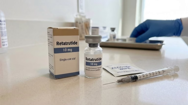
What Is RT10mg in Laboratory Research? Diagnostic and assay peptides RT10mg usually describes a 10 mg research presentation of Retatrutide.
The compound itself is Retatrutide / LY3437943 .
It is an engineered peptide.
Published research describes a 39-amino-acid sequence with lipid modification.
IUPHAR classifies Retatrutide as a triple peptidic agonist.
Its primary receptor targets are:
glucose-dependent insulinotropic polypeptide receptor, or GIPR; glucagon-like peptide-1 receptor, or GLP-1R; glucagon receptor, or GCGR. All three belong to the class B1 family of G protein-coupled receptors.
These receptors commonly signal through Gs proteins.
Consequently, intracellular cyclic AMP often provides an important experimental readout.
Why Researchers Study Three Receptors Separately Triple agonism does not mean all three receptors respond equally.
Ligand activity depends on receptor context.
Several variables can change the apparent response.
These include:
receptor density; cell background; assay temperature; incubation time; serum concentration; signaling amplification; detection technology. Therefore, one mixed cellular system may produce difficult-to-interpret data.
A cleaner design uses separate receptor-expressing systems first.
Researchers can then study combined biology later.
Retatrutide Receptor Architecture Structural biology has provided additional information about Retatrutide binding.
In 2024, Li and colleagues reported cryo-electron microscopy structures involving Retatrutide-bound GLP-1R, GIPR, and GCGR complexes.
Cryo-EM uses electron imaging to reconstruct macromolecular structures.
Therefore, researchers can directly examine peptide-receptor interactions.
These structures reveal both shared and receptor-specific contacts.
That information can guide:
receptor mutagenesis; ligand docking; structure-activity studies; signaling experiments; peptide analog design. However, a structural interaction does not directly equal functional potency.
Researchers still need cell-based assays.
The Core RT10mg Signaling Pathway The basic signaling model looks like this:
Retatrutide → receptor activation → Gs protein → adenylyl cyclase → cAMP → downstream signaling
This pathway applies broadly to GIPR, GLP-1R, and GCGR.
However, downstream biology can differ.
Cell context matters.
Therefore, cAMP represents a useful starting point rather than a complete biological description.
Designing a GIPR Assay A GIPR assay should isolate GIP receptor signaling.
One approach uses a cell line engineered to express human GIPR.
Researchers then expose the cells to a concentration series.
Next, they measure cAMP or another validated endpoint.
Important Controls A strong design may include:
vehicle control; native GIP reference ligand; positive assay control; RT10mg test material; background cells without GIPR where appropriate. The reference ligand confirms receptor responsiveness.
Meanwhile, background cells can reveal nonspecific effects.
Retatrutide discovery research reported substantial activity at human GIPR.
Therefore, GIPR provides an important component of the Retatrutide profile.
Designing a GLP-1R Assay GLP-1R represents another Retatrutide target.
Researchers can again use a receptor-expressing cell model.
However, assay conditions should remain controlled.
Useful comparison ligands may include native GLP-1 or a characterized GLP-1R agonist.
Semaglutide can also provide a mechanistic comparator in suitable research designs.
FDA documentation identifies semaglutide as a GLP-1 receptor agonist. Its reported molecular weight is 4113.58 Da.
By contrast, Retatrutide targets three receptor systems.
Therefore, the comparison can help isolate GLP-1R behavior from broader triple agonism.
Designing a GCGR Assay GCGR provides a major distinction between Retatrutide and dual GIP/GLP-1 agonists.
A GCGR-expressing cell model can isolate this pathway.
Researchers may use glucagon as a reference ligand.
Next, they can compare Retatrutide-mediated signaling.
Again, cAMP represents a logical endpoint.
However, researchers should validate assay sensitivity first.
This point matters because one concentration range may work well for GIPR but poorly for GCGR.
Therefore, researchers should not force all three receptors into one fixed concentration window without preliminary testing.
RT10mg vs Tirzepatide and Semaglutide in Receptor Assays Peptide Quality Control Retatrutide, tirzepatide, and semaglutide offer a useful mechanistic comparison.
Tirzepatide activates GIPR and GLP-1R.
Semaglutide primarily activates GLP-1R.
Retatrutide adds GCGR agonism.
This hierarchy can support a clean comparative experiment.
However, the compounds differ in sequence and molecular architecture.
Therefore, researchers should not interpret receptor count as the only variable.
Molecular Comparison for Experimental Planning Retatrutide has an approximate molecular mass of 4731.33 Da according to research reagent databases.
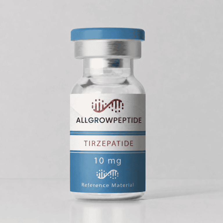
Tirzepatide has a molecular mass of 4813.53 Da .
Semaglutide has a molecular mass of 4113.58 Da .
This table illustrates why laboratories need compound-specific calibration.
Equal mass concentrations do not necessarily produce equal molar concentrations.
Therefore, researchers should calculate experimental concentrations using the appropriate molecular mass.
Why Molar Concentration Matters Peptide assays often compare compounds by molarity.
This approach compares molecule numbers more directly than mass concentration.
For example, 1 µg/mL of Retatrutide does not contain the same number of molecules as 1 µg/mL of semaglutide.
Their molecular weights differ.
Therefore, receptor pharmacology studies often use molar units.
This becomes especially important for concentration-response curves.
Researchers should document the molecular-weight value used for every calculation.
Building a Concentration-Response Experiment A concentration-response experiment examines how signaling changes across ligand concentrations.
Researchers generally need enough points to define:
baseline response; rising response; transition region; maximal or near-maximal response. However, one universal concentration range should not be copied between assays.
Receptor expression can shift apparent potency.
Likewise, detection technology changes assay sensitivity.
Therefore, laboratories should conduct a pilot experiment first.
The pilot can then guide the final concentration range.
EC50: Useful but Easy to Misinterpret The EC50 represents the concentration that produces half of the maximal measured response under defined conditions.
It provides a useful pharmacological parameter.
However, EC50 is not a fixed universal property.
It can change with:
receptor density; assay system; incubation time; signal amplification; cell type. Therefore, researchers should compare EC50 values only when experimental conditions are sufficiently similar.
This point becomes especially important for RT10mg.
The same Retatrutide preparation can produce different apparent EC50 values across GIPR, GLP-1R, and GCGR assays.
Practical Case: A Three-Receptor RT10mg Study Peptide Blends Consider a biotechnology laboratory studying receptor selectivity.
The researchers want to characterize one RT10mg batch.
They use three matched cell lines.
Cell System A: GIPR The first cell line expresses human GIPR.
Researchers include native GIP as a reference.
They measure cAMP.
Cell System B: GLP-1R The second line expresses human GLP-1R.
The team includes a characterized GLP-1R ligand.
Again, cAMP provides the primary endpoint.
Cell System C: GCGR The third line expresses human GCGR.
Glucagon serves as a receptor reference.
The team measures the same signaling endpoint.
Keep Experimental Variables Consistent The researchers use the same:
plate format; cell density strategy; incubation schedule; solvent system; detection platform; data-processing method. However, they validate the concentration range for each receptor.
This design improves interpretability.
A mixed cell system could make receptor attribution much harder.
Add a Negative Cell Control Receptor-negative cells can provide valuable information.
Suppose RT10mg produces a signal in cells lacking the target receptor.
That finding requires investigation.
Possible causes include:
endogenous receptor expression; assay interference; nonspecific cellular effects; solvent effects. Therefore, receptor-negative controls strengthen the experiment.
They become especially useful during assay development.
Why Receptor Expression Level Matters Two laboratories may use the same Retatrutide batch.
Yet their potency estimates may differ.
Receptor density represents one possible explanation.
Cells with high receptor expression can show greater signal amplification.
Therefore, receptor expression should remain reasonably stable between experiments.
Researchers can monitor expression through appropriate molecular methods.
Possible tools include:
qPCR; immunoblotting; flow cytometry; ligand-binding assays. Each method answers a different question.
For example, qPCR measures transcript abundance.
It does not directly quantify functional receptor at the cell surface.
Going Beyond cAMP cAMP provides only one view of GPCR biology.
Researchers can expand RT10mg studies with complementary endpoints.
β-Arrestin Recruitment β-arrestins participate in GPCR regulation.
Therefore, recruitment assays can reveal signaling behavior beyond G proteins.
Receptor Internalization Activated receptors may move away from the cell surface.
Researchers can track this process with imaging or labeled receptor systems.
Downstream Phosphorylation Some experiments measure downstream kinase pathways.
However, these responses may involve several signaling networks.
Therefore, interpretation becomes more complex.
Transcriptomic Analysis RNA sequencing can examine broader cellular responses.
Yet transcriptional changes occur far downstream of receptor binding.
Researchers should therefore combine them with direct receptor assays.
Structural Biology Can Guide Mutagenesis The 2024 structural study provides cryo-EM views of Retatrutide interactions with its three receptor targets.
Researchers can use this information to select receptor residues for mutagenesis.
For example, a laboratory may identify a contact residue.
Next, the team creates a receptor mutant.
Then it compares Retatrutide signaling with wild-type receptor signaling.
However, reduced activity alone does not prove a direct binding effect.
The mutation may reduce receptor expression.
Therefore, expression controls remain necessary.
This illustrates an important experimental principle:
Structural evidence and functional evidence should support each other.
RT10mg Quality Control Before Functional Assays Functional experiments require chemically defined material.
Therefore, receptor research should start with sample verification.
HPLC HPLC can assess chromatographic purity.
However, it cannot independently confirm identity.
LC-MS LC-MS provides molecular-mass information.
Therefore, it strengthens identity verification.
COA The Certificate of Analysis should connect the analytical results with the actual batch.
The RT10mg product page on ALLGROW already emphasizes batch-specific documentation and mass-spectrometry information where available.
Researchers should confirm these records before initiating quantitative assays.
Controlling Batch Effects Long experiments may require several RT10mg lots.
However, switching batches can introduce variability.
Therefore, researchers should consider a bridge experiment.
Use the old and new lots in one assay run.
Keep all other conditions constant.
Then compare predefined assay metrics.
Useful parameters may include:
curve shape; maximum response; apparent potency; baseline behavior. Researchers should establish acceptance criteria in advance.
This reduces post-hoc interpretation.
RT10mg Storage During Cell-Based Research The ideal storage condition depends on formulation and validation data.
Therefore, researchers should follow lot-specific documentation.
Lyophilized peptide should remain protected from unnecessary moisture.
In addition, researchers should minimize temperature cycling.
Once a research solution has been prepared, several variables become more important.
These include:
buffer composition; pH; protein content; container surface; temperature; storage duration. Repeated freeze-thaw cycles may add variability.
Consequently, working aliquots can help in multi-day experiments.
However, laboratories should validate their own protocol.
Published Retatrutide Research Context Retatrutide has moved through an extensive clinical development program.
The original molecular research established its GIPR, GLP-1R, and GCGR agonist profile.
ClinicalTrials.gov lists TRIUMPH-1 as a Phase 3 Retatrutide study in participants with obesity or overweight.
Lilly reported top-line TRIUMPH-1 results in May 2026.
The trial randomized 2,339 participants across Retatrutide and placebo groups.
At 80 weeks, Lilly reported mean body-weight changes of −19.0%, −25.9%, and −28.3% for the 4, 9, and 12 mg groups under the efficacy estimand.
These figures provide clinical-development context only.
They are not specifications for an RT10mg research vial.
They also should not guide laboratory material dosing.
Retatrutide remains investigational as of August 2026.
Common Experimental Mistakes in RT10mg Research Mistake 1: Using One Cell Line to Claim Triple-Receptor Selectivity One cell line may express several endogenous receptors.
Therefore, receptor-specific systems provide clearer data.
Mistake 2: Comparing Mass Concentrations Directly Different peptides have different molecular weights.
Use molar comparisons when the research question requires them.
Mistake 3: Ignoring Batch Identity A label cannot replace HPLC and MS documentation.
Mistake 4: Comparing EC50 Values Across Different Assays Assay conditions strongly influence apparent potency.
Therefore, cross-study comparisons require caution.
Mistake 5: Treating cAMP as the Entire Mechanism cAMP provides important information.
However, GPCR regulation also involves receptor trafficking and other signaling pathways.
Frequently Asked Questions About RT10mg Research What is RT10mg? RT10mg commonly refers to a 10 mg research presentation of Retatrutide. The scientific identifier is LY3437943. Retatrutide is a modified peptide agonist that targets GIPR, GLP-1R, and GCGR.
Is RT10mg a peptide? Yes. Retatrutide is a peptide molecule. Published research describes a 39-amino-acid structure with a C20 fatty diacid modification.
Which receptors does Retatrutide activate? Retatrutide activates GIPR, GLP-1R, and GCGR. Therefore, researchers classify it as a triple receptor agonist.
Why is cAMP measured in Retatrutide research? All three primary receptor targets can signal through Gs proteins. Gs activates adenylyl cyclase, which increases intracellular cAMP. Therefore, cAMP provides a useful receptor-function endpoint.
Can the same RT10mg concentration range be used for all three receptors? Not automatically. Apparent potency can differ by receptor and assay system. Therefore, researchers should validate the range separately for GIPR, GLP-1R, and GCGR.
What is the difference between RT10mg and tirzepatide? Retatrutide targets GIPR, GLP-1R, and GCGR. Tirzepatide targets GIPR and GLP-1R. FDA documentation reports tirzepatide as a dual receptor agonist.
What is the difference between RT10mg and semaglutide? Semaglutide primarily targets GLP-1R. Retatrutide targets three receptor systems. Their molecular weights also differ. FDA reports semaglutide at 4113.58 Da.
Can Retatrutide and tirzepatide appear in the same experiment? Yes, when the research question requires comparison. However, researchers should control receptor system, molar concentration, purity, and assay conditions.
How should RT10mg identity be confirmed? LC-MS provides useful molecular-mass evidence. HPLC adds chromatographic purity information. Therefore, the two methods provide complementary evidence.
Why should researchers use receptor-negative cells? They help detect nonspecific signaling or endogenous receptor effects. Therefore, negative cell controls improve receptor-attribution confidence.
Does an RT10mg COA prove biological potency? No. A COA may document identity and chemical purity. Functional potency requires a suitable biological assay.
Is Retatrutide approved? No. Retatrutide remains an investigational molecule as of August 2026. Lilly continues to identify it as an investigational triple hormone receptor agonist.
Conclusion RT10mg research provides a useful framework for studying multi-receptor peptide pharmacology.
Retatrutide targets GIPR, GLP-1R, and GCGR within one engineered peptide. Therefore, it can support receptor-selectivity and signaling research.
However, triple agonism also increases experimental complexity.
Researchers should isolate receptor pathways where possible.
They should also control cell background, receptor expression, concentration units, batch identity, and analytical quality.
Moreover, cAMP data should not stand alone when the scientific question extends to receptor trafficking or signaling bias.
Combining chemical verification with controlled functional assays creates stronger evidence.
That approach also makes RT10mg experiments easier to reproduce.
For Research Use Only. Not for human use.
References Coskun T, et al. LY3437943, a novel triple glucagon, GIP, and GLP-1 receptor agonist. Cell Metabolism. 2022. Li W, et al. Structural insights into the triple agonism at GLP-1R, GIPR and GCGR manifested by retatrutide. Cell Discovery. 2024;10:77. IUPHAR/BPS Guide to Pharmacology. Retatrutide / LY3437943. FDA. Mounjaro (tirzepatide) prescribing information. Molecular structure and GIPR/GLP-1R mechanism. FDA. Semaglutide prescribing information. Molecular structure and GLP-1 receptor mechanism. ClinicalTrials.gov. TRIUMPH-1, NCT05929066. Eli Lilly and Company. TRIUMPH-1 Phase 3 top-line results. May 21, 2026.